List The Main Structures Of The Eye
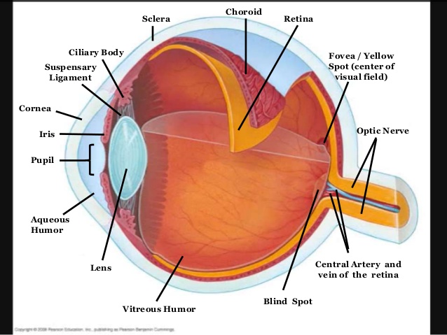
Why is it called the blind spot. It is the eyes primary light-focusing structure.
 Activity Sheet 1 How The Eyes Work Human Eye Diagram Human Body Activities Teaching Biology
The retina is the innermost layer of the eye.

List the main structures of the eye. The eyes are formed from several embryonic layers. The main muscles of the eye are Lateral rectus Medial rectus Superior rectus and inferior rectus. The neuroectoderm produces the posterior part of the iris optic nerve and retina.
The epithelium of the cornea and lens are derived from the surface ectoderm. The basic function of these muscles is to provide different tensions and torques that further control the movement of the eye. The sclera comprises the majority of the fibrous layer approximately 85.
There are two main types of photoreceptors. The eye has many parts which work together to accomplish vision and to keep the structures required for vision safe from infection and injury. The eye consists of three layers of tissue which make up the wall of the eye.
The outer transparent structure at the front of the eye that covers the iris pupil and anterior chamber. It includes the medial rectus lateral rectus superior rectus inferior rectus inferior oblique and superior oblique. The surface of the eye and of the inner eyelids is covered by a clear protective membrane called the conjunctiva.
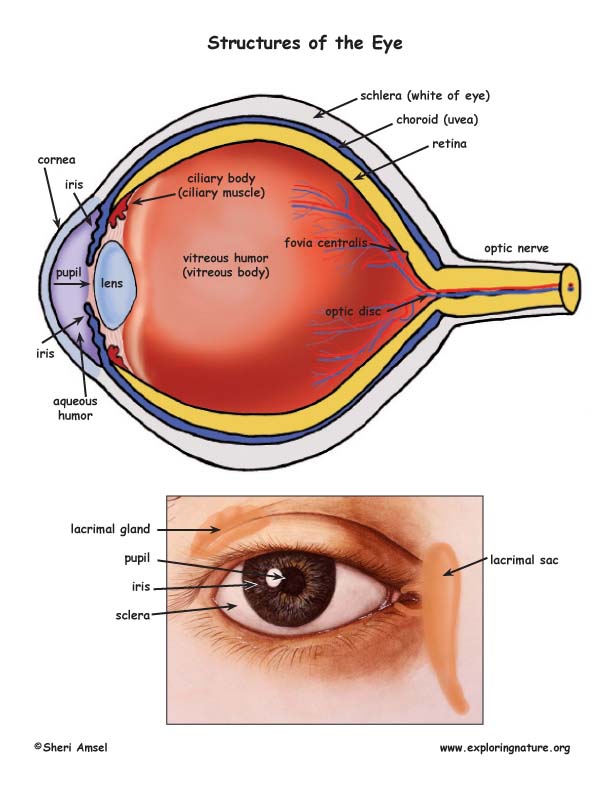
If too steep myopianearsightedness. The main parts of the human eye are the cornea iris pupil aqueous humor lens vitreous humor retina and optic nerve. Choroid Cornea Fovea Iris Macula Lens Optic Nerve Pupil Retina Sclera Vitreous Humor.
This layer is a very stable fibrous membrane that continues to retain the shape of the eye and provides protection. The blind spot has the region on retina which is known as the optic disk. Light enters the eye by passing through the transparent cornea and aqueous humor.
Causes loss of central vision as you get older. The Cornea is the second structure that light strikes. The dome-shaped layer protects human eye from elements against entering in the inner parts of the eye.
The endothelium of the cornea sclera and choroid arise from the neural crest cells. An eye also consists of six muscles. Eyeball Bulbus oculi The eye is a highly specialized sensory organ located within the bony orbitThe main function of the eye is to detect the visual stimuli photoreception and to convey the gathered information to the brain via the optic nerve CN IIIn the brain the information from the eye is processed and ultimately translated into an image.
The sclera is the outermost layer of tissue also called the white of the eye. The conjunctiva is the membrane that lines the eyelids and covers the white of the eye and the cornea is the clear layer in front of the iris and pupil. Their main functions are to provide shape to the eye and support the deeper structures.
It is the clear transparent front part of the eye that covers the iris pupil and anterior chamber and provides most of an eyes optical power if too flat hyperopiafarsightedness. It needs to be smooth round clear and tough. It consists of the sclera and cornea which are continuous with each other.
Deposits of yellowish extra cellular waste products that accumulate within and beneath the retinal pigmented epithelium RPE layer. It houses more than 120 million light-sensitive photoreceptor cells that detect light and convert it into electrical signals. Here are the main Eye Parts of human eye by which a human can see around himself.
View this anatomical diagram of the eye showing the eye structure including the pupil iris cornea retina and optic nerve. Anatomically the eye comprises two components fused into one. In what area of the eye is the blind spot located.
The fibrous layer of the eye is the outermost layer. Keratoconjunctivitis sicca is dryness of the conjunctiva and cornea. The central artery supplies the retina while the central vein drains the retina.
There are many other layers of cornea that provide more protection. The cornea is the outer layer covering of the eye. Hence it does not possess a perfect spherical shape.
Central Artery and Vein The central artery and vein runs through the center of the optic nerve. External components include structures which can be seen on the exterior of the eye and internal components include structures present within. Cones are responsible for sharp detailed central vision and color vision and are clustered mainly in the macula.
Often called lazy eye this condition starts in childhoodOne eye sees better than the. These signals are sent on. How the eye works and descriptions and functions of the major structures of the human eye.
Rods are responsible for night and peripheral side vision.
 Vision And The Structure Of The Eye
 The Camera Approach For Developing Functional Bionic Eyes Bionic Eye Bionic Eyes
 This Video Summarizes The Eye It Focuses On How Structures Work Together To Refract Light In Order To Focus On The Reti Eye Anatomy Human Eye Parts Of The Eye
 Anatomy Of The Eye Human Eye Anatomy In 2020 Eye Anatomy Human Eyeball Human Eye
 Eye Anatomy Functions And Structure Eye Anatomy Senses Anatomy
 There Are Three Layers Or Tunics Of The Eyeball The Fibrous Layer Is The Outermost Layer That Contains The Medical Knowledge Medical School Studying Midterm
 Anatomy Of The Internal Eye Sagittal Views Depict A The Three Tunics Of The Eye And B Internal Eye Structures Eye Anatomy Anatomy Eyes
 Free Anatomy Coloring Pages Homeschool Giveaways Anatomy Coloring Book Human Eye Eye Anatomy
 Human Eye Human Eye Diagram Human Eye Eyes
 Do You Know Which Part Of Your Eye Is Which Are You Kids Very Curious About Eyes Check Out This Article To Learn M Diagram Of The Eye Articles For Kids
 Eye Anatomy Diagram Illustrations 41 New Ideas Eye Anatomy Eye Anatomy Diagram Anatomy
 Sclera White Of The Eye Medical Anatomy Eye Structure Human Eye Diagram
 Iris The Sacred Meeting Place Between Science And Spirituality Iridology Chart Iridology Eye Chart
 Pin On The Eyes Have It
 What Are The Three Layers Of The Human Eye Socratic
 Pin On Eye
 Human Eye The Visual Process The First Line Of Protection Of The Eyes Is Provided By The Lids Which Prevent Access Of Forei Body Anatomy Cow Eyes Human Eye
Anatomy Of The Eye Hot Air Balooning Eye Anatomy Parts Of The Eye Anatomy
 Pin On Weapon And War Anatomy
Post a Comment for "List The Main Structures Of The Eye"