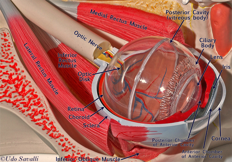
Structure Of Eye With Labelling
The cornea is a clear covering that protects the front of the eye and in the back the optic nerve is responsible for sending electrical signals to the brain. The eyes are a very complex organ and so there are many named parts to remember each with their own specific function.
 The Human Eye Labelling Activity
Images and pdfs - Just in case you get tired of looking at the screen weve provided images and pdf files that you can print out and use for off-line practice.
Structure of eye with labelling. Hence it does not possess a perfect spherical shape. Label Parts of the Human Eye. This quiz has tags.
Lens - a crystalline structure located just behind the iris - it focuses light onto the retina. From the cornea to the optic nerve and every part in between this science quiz game will help make you an expert on the parts of the human eye. The human eye is a roughly spherical organ responsible for perceiving visual stimuli.
It is enclosed within the eye sockets in the skull and is anchored down by muscles within the sockets. Test your knowledge on this science quiz and compare your score to others. In the class today we covered parts of the eye and what changes in them should be alarming to a patient.
From the quiz author. Word Roots - When you learn the word roots prefixes and suffixes contained within anatomical and medical terms you can often work out what they mean. Take up this quiz and find out.
People say that the eyes are the windows to a persons soul. The image is taken from above the left eye. The white of your eye.
How much did you get to understand about the human eye. The colour of the iris actually indicates the colour of the eye. When showing this human eye labelling parts our team can guarantee to impress you.
Iris optic nerve pupil cornea lens retina. What are the parts of the eye. People say that the eyes are the windows to a persons soul.
Visit an eye care unit or clinic with your friends and teacher to observe various charts displaying the anatomy of eye. Light enters the eye by passing through the transparent cornea and aqueous humor. Label the parts of the human eye as quickly as possible.
The iris controls the size of the pupil which is the opening that allows light to enter the lens. 3 DIABETES AND HEALTHY EYES ANATOMY OF THE EYE AND ITS FUNCTION Vision is wonderful but you could lose it if you have diabetes. There are 7 bones of the orbit two groups of muscles intrinsic ocular and extraocular three layers to the eyeball and thats just the beginning.
Just behind the iris. Anatomically the eye comprises two components fused into one. Nearsighted - also called myopia a condition in which nearby objects are seen more clearly than distant objects because light is focused in front of the the retina not on it.
Check Your Progress B. There is a printable worksheet available for download here so you can take the quiz with pen and paper. When light shines on an object a reflection is sent which passes through the eye lens and later projects the image of the object on the retina.
Select One Anterior Chamber Ciliary Body Cornea Fibrous Tunic Iris Lateral Rectus Muscle Lens Medial Rectus Muscle Optic Disk Optic Nerve Pupil Retina Vascular Tunic Vitreous Nerve. Displaying top 8 worksheets found for - Label The Parts Of The Eye. 2015 WebMD LLC.
FIeld of VISIon And dynAmIc rAnge of HumAn eye. For us to see there has to be light. Label the parts of the eye in Figure 12 and also list them in the Table given below.
Further refracts light to focus it onto the retina. Quiz created specifically to line up with MillerLevine Biology textbook ISBN 0131152912. The anatomy and physiology of the human eye is an important part of many courses eg.
Heres a list of the main ones. Some of the worksheets for this concept are The human eye Teachers guide vision grades 3 to 5 Memento mer exhibit and See well for a lifetime parts of the eye Nose eye ear hand arm head Teachers guide hearing grades 3 to 5 Label the ear diagram for kids 3 side view 7. Youve learned what the parts of the eye are and labelled them on the sheet so now find out what they do for our vision.
The iris also helps regulate or adjust exposure by adjusting the iris. Controls how much light enters the pupil. The main parts of the human eye are the cornea iris pupil aqueous humor lens vitreous humor retina and optic nerve.
A dark muscular tissue and ring-like structure behind the cornea are known as the iris. Can you locate the parts of the human eye. Healthy Eyes Toolkit Parts of the Eye To understand eye problems it is helpful to know the different parts of the eye.
9th and 10th grade Texas Biology. How to learn the parts of the eye Found within two cavities in the skull known as the orbits the eyes are surrounded by several supporting structures including muscles vessels and nerves. This is an online quiz called Label the Eye.
Please refer to the back of this handout for descriptions of their functions. This is an exercise for students to label a simple blank eye diagram with the following parts. Refracts light - bends it as it enters the eye.
A thin layer of tissue that covers the entire front of your eye except for the cornea. The main parts of. Light enters the eye through the cornea.
 Simple Eye Diagrams Easy Eye Diagram Labeled Eye Diagram Human Eye Diagram Eye Structure Simple Eye
 Module 1 Labeled Diagram Of The Eye Diagram Of The Eye Dot Worksheets Eye Anatomy
 Eye Anatomy Diagram Enchantedlearning Com Eye Anatomy Anatomy Eye Anatomy Diagram
 Simple Eye Diagrams Easy Eye Diagram Labeled Eye Diagram Human Eye Diagram Medicine Images Simple Eye
 The Miracles In Our Body Eye Anatomy Eye Anatomy Diagram Human Anatomy And Physiology
 Human Eye Anatomy In Detail Www Anatomynote Com Ear Anatomy Eye Anatomy Anatomy And Physiology
 Pin On Vision
 Anatomy Eye Diagram To Label Eye Anatomy Diagram Eye Anatomy Anatomy
 Simple Eye Diagrams Easy Eye Diagram Labeled Eye Diagram In 2020 Simple Eye Human Eye Diagram Eye Structure
 Human Eye Anatomy Parts Of The Eye Explained Eye Anatomy Anatomy Human Anatomy And Physiology
 The Eye Diagram For Kids Human Eye Diagram Science Anchor Charts Fun Science
 Aao G04 Large Jpg 1320 1020 Human Eye Diagram Eye Anatomy Human Anatomy Drawing
 Worksheet With Answer For Structure Of Human Eye Anatomy Coloring Book Eye Anatomy Human Body Unit
 Pictures Of Human Heart Anatomy Anatomy Of The Human Heart 4k Ultra Hd Wallpaper Human Heart Anatomy Human Anatomy And Physiology Heart Anatomy
 Eyes Layers Of Learning Human Eye Diagram Parts Of The Eye Human Eye
 Human Body Organs Diagram From The Back Koibana Info Human Body Organs Body Organs Diagram Human Body Anatomy
 Eye Diagram By Firkin Human Eye Diagram Diagram Of The Eye Eye Structure
 Diagram Of Basic Parts Of The Eye Eye Anatomy Parts Of The Eye Diagram Of The Eye
Post a Comment for "Structure Of Eye With Labelling"