Eye Anatomy Tear Duct
The lacrimal gland lacrimal sac and nasolacrimal duct. Further components of the tear film include an inner mucous layer produced by specialized Read More.
 Pin On Skeleton Anatomy
Tears consist of a complex and usually clear fluid that is diffused between the eye and the eyelid.

Eye anatomy tear duct. Gross anatomy the nasolacrimal duct is the inferior continuation of the lacrimal sac and is 17 mm in length in total. The nasolacrimal duct also called the tear duct carries tears from the lacrimal sac of the eye into the nasal cavity. The duct begins in the eye socket between the maxillary and lacrimal bones from where it passes downwards and backwardsThe opening of the nasolacrimal duct into the inferior nasal meatus of the nasal cavity is partially covered by a mucosal fold valve of Hasner or plica.
Tear drains from the eyes in to the nose through the tear duct. For ophthalmologists optometrists medical dental and optometry students eye-anatomy forms the basis for eye-pathology in diseases. The tear duct is part of the tear drainage system.
The tears flow down the surface of your eye to lubricate and protect. The duct begins in the eye socket between the maxillary and lacrimal bones from where it passes downwards and backwards. A blocked tear duct is when the eyes drainage system for tears is either partially or completely obstructed.
The second layer of the tear film is the aqueous layer and is secreted by the lacrimal glands which are located just below the eyebrow on the temporal near the temple side of the eyelid. Eye drops and ointments are usually prescribed first for blocked tear ducts. The tear duct also called the nasolacrimal duct is located in the inner corner of the eye and is part of the tear drainage system that goes from the eye through the back of the nose and down the throat.
Dry eye retinal detachment. The nasolacrimal drainage system serves as a conduit for tear flow from the external eye to the nasal cavity. This is why a teary eye is usually accompanied by a runny nose.
Anatomy of the human eye Anatomy of the eye includes lacrimal gland cornea conjunctiva uvea iris choroid ciliary body lens blood supply retina vitreous optic-nerve. However if the tear ducts block then tears have no option but to spill over the face. In unusual cases obstructed tear ducts may be brought on by a tumor.
To use the eye drops shake the bottle well tilt your head back then place the recommended number of drops into the eye. According to MSD Vet the symptoms of a blocked tear duct are mainly wet fur beneath the eye. It consists of the puncta canaliculi lacrimal sac and nasolacrimal duct see the.
The tear duct is also called the nasola. Tears cannot drain normally causing a watery irritated or chronically infected eye. In tear duct and glands lachrymal or lacrimal duct and glands structures that produce and distribute the watery component of the tear film.
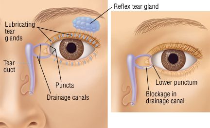
Dacryocystitis refers to an infection of the tear sacs which are part of the tear drainage system in the eye. Tear glands and tear ducts The tear glands lacrimal glands located above each eyeball continuously supply tear fluid thats wiped across the surface of your eye each time you blink your eyelids. Tears drain from each eye through small canals drainage canals a tear sac and a tear duct.
Tears consist of a complex and usually clear fluid that is diffused between the eye and the eyelid. The sebaceous meiboman glands are also seen in the diagram on the left. It drains tears through the nasal bone and into the back of the nose.
Tear duct and glands also called lachrymal or lacrimal duct and glands structures that produce and distribute the watery component of the tear film. This is a nice anatomical detail Mother Nature added to eyes to avoid a permanently wet face. Most of your tears come from your lacrimal glands which are located above each eye.
Close the eye for 30 seconds to a minute to allow the eye drops to be absorbed. The tears are spread across the eyes surface to moisten clean nourish and protect the ocular tissues. You will need to visit your doctor to get a prescription.
When a person blinks it spreads their tears over the surface of their eye. Abbs on March 29 2020. The tear duct is part of the tear drainage system.
On January 14 2020. And tear duct obstruction is often a side effect of chemotherapy treatments for cancer. Excess fluid drains through the tear ducts into the nose.
Drainage canals are found at the inner corner of each upper and lower eyelid and they carry away tears that have rinsed the front surface of the eye. The process begins in the lacrimal glands which are located in the outer upper corner eye socket on each side of the eye. The job of the duct is to drain away excessive amounts of tear fluid.
This is a small tube that runs from the eye to the nasal cavity. THANKS SO MUCH. This is really helpful with my information reports.
Tear ducts are part of the nasolacrimal system which is responsible for draining tears from the surface of the eye. Nasolacrimal duct the nasolacrimal duct also called tear duct latin. An injury to the eye or nose can interrupt the tear ducts function and even something as little as dust or dirt stuck in the tear duct can cause problems.
 Lacrimal Apparatus Lacrimal Gland Lacrimal Canaliculi Lacrimal Sac Nasolacrimal Duct Lacrimal Apparatus A System Custom Contact Lenses Eye Care Eye Doctor
 Tearing Landa Landa Eye Care Specialists Skeletal System Activities Tears In Eyes Eye Care
 Lacrimal Apparatus Anatomy Eye Exercises Eye Health Eye Anatomy
 Lacrimal Gland Anatomy Human Anatomy And Physiology Aesthetic Medicine Medical Anatomy
 Konnycsatorna Dry Eyes Swollen Eyelids Remedy Dry Eyelids
 Tear Duct Infection Landa Landa Eye Care Specialists Llc Tearing Anatomie Korper Anatomie Augen Krankheit
 Lacrimal Anatomy Of The Eye Human Anatomy And Physiology Medical Anatomy Aesthetic Medicine
 Tear Duct Obstruction And Surgery Eye Anatomy Anatomy And Physiology Eye Facts
 Diagram Illustrating Blocked Tear Ducts In The Human Eye Human Eye Eye Illustration Blocked Tear Duct
 Blocked Tear Duct Symptoms And Causes Mayo Clinic In 2021 Dry Eye Symptoms Dry Eyes Dry Eye Syndrome
 Lacrimal Apparatus The Lacrimal Apparatus Continually Produces Lacrimal Fluid Tears That Cleanses And Maintains A Moist Condit Nasal Cavity Cavities Anatomy
Mr David Cheung Consultant Ophthalmologist Oculoplastic And Orbital Surgeon Specialising In Functional Reconstru Blocked Tear Duct Eyelid Lift Tear Trough
 Blocked Tear Duct In Adults Blocked Tear Duct Duct Eye Infection Symptoms
 Tear Duct Lacrimal Tearing Tearduct Tear Duct System Description Of Blockage Duct Dry Eye Treatment Dry Eye Syndrome
 Image Result For Blocked Tear Duct Pictures Blocked Tear Duct Duct Basic
 Tear Duct Infection Dacryocystitis Harvard Health Eye Infection Symptoms Eye Infections Tears
 Lacrimal Apparatus Includes Lacrimal Sac Gland Punctum Canaliculus Nasolacrimal Duct Inferior Meatus Of Nasal Cavity Jpg 580 Eye Skin Care Optometry Eye Facts
 Valve Of Hasner In Nasolacrimal Duct Lacrimal Inferior Canaliculus Involved More Commonly In Canalicular Lacerations Medical Memes Duct Medical Studies
 Related Image Eye Drawing Eye Drawing Tutorials Realistic Eye Drawing
Post a Comment for "Eye Anatomy Tear Duct"