Structure And Function Of Eye And Ear
Eye Structure and Function. The ear is divided into three parts.
 Contentseyesvideo Anatomy And Function Of The Eyeearsvideo Ear Anatomy Our Most Important Sensory Recept Ear Anatomy Eye Anatomy Human Anatomy And Physiology
Functions of External Ear.

Structure and function of eye and ear. Spiral-shaped fluid filled area of the ear which is comprised with tiny hairs that react to signs of vibration. Eardrum Thin membrane the reacts to vibration impulses to invoke a physical response to the rest of the ear. It directs sound waves towards the tympanic membrane.
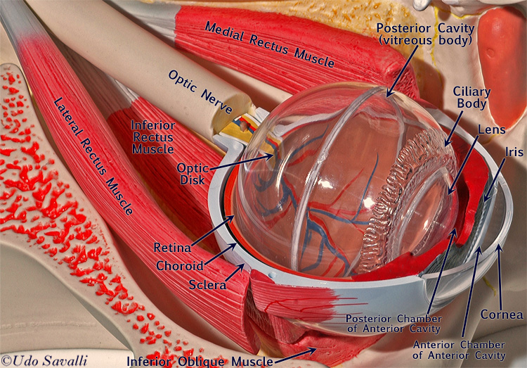
The outer ear consists of the visible portion called the auricle or pinna which projects from the side of the head and the short external auditory canal the inner end of which is closed by the tympanic membrane commonly called the eardrum. The Structure and Function of the Human Eye The human eye is essentially a three layered structure comprising an outer fibrous layer the sclera and cornea a middle vascular layer the choroid ciliary body and iris and inner retina neurons and photoreceptor cells. The innermost is the retina which gets its circulation from the vessels of the choroid as well as the retinal vessels which can be seen in an ophthalmoscope.
Both are located in the front part of the eye in front of the lens. Components The eye is made up of three coats enclosing three transparent structures. The malleus transmits sound vibrations from the eardrums to the incus.
The eye receives oxygen through the aqueous. It bends or refracts light. The structure of eye comprises three coats within which further are three transparent structures.
Ring of muscle around the pupil that controls amount of light that enters. It is the pigmented coloured portion of the eye visible externally. Structure and function of eye 1.
The outer ear is composed of the auricle pinna and the external auditory canal ear canal Sisco. Blood vessels can be seen within the sclera as well as a. The outermost layer or the fibrous tunic consists of the cornea and sclera.
Call lens has a lens that is adjustable according to the distance called a diaphragm pupil whose diameter is regulated by the iris and a sensitive tissue light which is the retina. The ears are the organs of hearing and equilibrium. The outer ear functions to catch sound waves and funnels them through the ear canal to the middle ear Inner body.
Light enters through the cornea the transparent outer covering of the eye. In terms of the former the outer ear is shaped to direct sound waves from the external environment to the ear canal. The main function of the iris is to control the diameter of the pupil according to the light source.
The main function is to refract the light along with the lens. The round window function of human ear is to keep the cochlear fluids contained within the scala vestibuli and scala tympani. The external the middle and the inner parts.
Acoustic energy in the form of sound waves passes pinna ear canal. These are then directed towards the tympanic membrane eardrum causing it to vibrate. Its function is to nourish the cornea iris and lens by carrying nutrients it removes waste products excreted from the lens and maintain intraocular pressure and thus maintains the shape of the eye.
Structure and Function of Eye and Ear Structure and Function of Eye and Ear Structure and Functions of the Eye The human eye works very similar to that of most vertebrates and some mollusks. Nerve that carries impulses from the eye to be interpreted by the brain into images. The sound waves produce pressure changes over the surface of the tympanic membrane.
Sight and sound are two of the most beautiful senses we possess. By sensing the movements of these stones the ear can tell our brain where we are relative to the directions up and down and how our body is moving or accelerating. These middle ear bones mechanically amplify sound and compensate mismatched impedance.
It is the small aperture located in the centre of the Iris. Back layer of the eye made up of light sensitive cells. For this reason it is quite important to comprehend the structure of eye and ear accompanied by their functions.
The inner ear is an incredibly complex helical structure formedwithin the skull wall. It operates mechanically driven by the middleear hydrodynamically by vibrations through fluid-filled chambersand electrochemically by the trading of ions between two separatelycharged fluids. To understand how the eye sees it helps to know the eye structures and functions.
The outermost layer known as the fibrous tunic is composed of the cornea and sclera. Distinguishes light and darkshape colour brightness distance ofobjects. Anatomically the ear has three distinguishable parts.
The function of the outer ear is to collect sound waves and guide them to the tympanic membrane. The cerumen ear wax prevents the entry of the foreign bodies into the ear. The middle layer known as the vascular tunic or uvea consists of the choroid ciliary body and iris.
Primarily the ear serves two functionshearing and regulation of balance. A sound wave also known as a pressure wave is a repeating pattern of high pressure and low pressure regions moving through a medium Henderson. Gift of the Creator Gives us the sense of sight 70 of all sensory receptors are in the eye Spheroid structure about 23 the size of aping-pong ball Functions.
This gives the eye its shape. It also functions as a multiplier of the sound waves generated from the oval window membrane. Die off as we age or to the constant presence of loud noise.
In humans the inner ear contains parts called the semicircular canals where otoliths tiny stone-like structures shift in response to gravity and the movement of our body. The outer middle and inner ear. Sound waves hit the ear drum causing it to vibrate like a drum.
The external ear is the outer funnel-like structure called the auricle or pinna and the external auditory meatus is called the external auditory canal. Transparent convex device in the eye that refracts light to create an image. The eyeball is rounded so the cornea acts as a lens.
It sets three ossicle bones malleus incus stapes into motion changing acoustic energy to mechanical energy.
 Special Senses Anatomy And Physiology Nurseslabs Ear Anatomy Anatomy And Physiology Physiology
 The Ear Diagram And Functions Human Ear Diagram Human Ear Human Anatomy And Physiology
 Aao G04 Large Jpg 1320 1020 Human Eye Diagram Eye Anatomy Human Anatomy Drawing
 Eye And Ear Models Eye Anatomy Anatomy Models Medical Anatomy
 Sound Waves Structure Of The Ear Have Your Students Label A Diagram Of A Human Ear Listing The Different Functions O Human Ear Diagram Sound Waves Ear Parts
 Total 0 Average 0 5 Human Ear Structure And Function A Structure Of Human Ear The Ear Consists Of Three Comp Ear Structure Human Ear Diagram Human Ear
 The Anatomy Of The Ear Infographic Basic Anatomy And Physiology Ear Function Ear Anatomy
 Human Ear Anatomy Pics Human Ear Anatomy Ear Anatomy Ear Structure
 The Science Of Eye Health Exercises For Injuries Eye Anatomy Human Eye Diagram Eye Anatomy Diagram
 Human Ear Anatomy Ears Inner Structure Organ Of Hearing Vector Illus By Tartila Thehungryjpeg Com Ears Affiliate Human Ear Anatomy Ear Anatomy Human Ear
Functions Of The Parts Of The Eye Parts Of The Eye Eye Anatomy Anatomy And Physiology
 Human Ear Home Tuition Guwahati Assam Ear Diagram Ear Anatomy Outer Ear
 Eyeballzzz Eye Anatomy Anatomy Human Anatomy Chart
 Here S How The Human Eye Works Human Eye Human Eye Diagram Eye Structure
 Pin On Learn Anatomy Human
 The Parts Of The Eye External Ear Anatomy Ear Anatomy Anatomy
 Labelled Diagram Of The Ear Ear Diagram Ear Anatomy Ear Structure
 Labeled Eye Diagram Human Eye Diagram Eye Anatomy Diagram Of The Eye
Post a Comment for "Structure And Function Of Eye And Ear"