Ct Anatomy Of Brain
Ct head neck atlas. HISTORY Sir Godfrey hounsfield-1972 Nobel prize in 1979 with cormack six generation of scanners Latest 128 multidetector ct 3.
 Imaging Acute Stroke Youtube Make It Simple Easy Easy Youtube
What is a CT scan of the brain.

Ct anatomy of brain. 3- Superior Sagittal Sinus. 2- Parietal Lobe of Cerebrum. Brain bones of cranium sinuses of the face.
6 frontal bone 27 occipital bone 32 optic nerve 37 basilar artery. Third and fourth ventricles in midline. The temporal lobes occupy the middle cranial fossae.
Head CT Approach First - evaluate normal anatomical structures window for optimal brain tissue contrast Second assess for signs of underlying pathology such as. Brain lobes - CT brain inferior slice The most anterior parts of the frontal lobes occupy the anterior cranial fossae. 5- Body of Lateral Ventricle.
Its hemispheric surface is convoluted and has gyri and sulci. The anterior part of the head is at the top of the image. To load the neck ct anatomy module in a new window click on its image above.
The brain consists of the cerebrum cerebellum and brainstem. Ct brain basics and anatomy 1. Pituitary and pineal glands are regrouped as gland.
CT brain - image orientation. CT BRAIN-BASICS AND ANATOMY M3 BRAINSTORMING 231109 2. Basic radiological anatomy of the brain and spine with annotated CT and MRI images covering the brain including the brainstem structures and ventricles and whole spine.
The study of the vascularisation of the brain is possible with the arteries and venous sinuses sections. Non contrast axial ct head. 55- Occipital Lobe of Cerebrum.
Anatomy ct axial brain form no 19. CT images of the brain are conventionally viewed from below as if looking up into the top of the head. Also called ambient cistern is a cistern of the subarachnoid space between the posterior end of the corpus callosum and the superior surface of the cerebellum.
Through cerebral CT radiographers see the patients brain without the need for surgery. Head CT Scan Intracranial CT Scan A CT of the brain is a noninvasive diagnostic imaging procedure that uses special X-rays measurements to produce horizontal or axial images often called slices of the brain. Mass effect edema midline shift hemorrhage hydrocephalus subdural or epidural collectionhematoma or infarction Third evaluate sinuses and osseous structures.
Cerebral lobes Basal ganglia Lentiform nuclei Thalamus Vascular territories Internal capsule calcifications fossa posterior. Spl head and neck atlas 2012 november. Anatomy CT Axial Brain Form No 4.
Lateral ventricles of normal volume. Overview of Brain Anatomy The brain is semisolid and conforms to the shape of the skull. As the cursor is moved over a particular anatomical area that area is highlighted and labeled.
Anatomy of the head on a cranial ct scan. Brainstem and cerebellum without evidence of focal lesions. Brain CT scans can provide more detailed information about brain tissue and brain structures than standard X-rays of the head thus providing more.
CT anatomy of the brain. Neck ct cervical lymph node levels. 5- Body of Lateral Ventricle.
Cerebral Computed Tomography CT Cerebral computed tomography CT is a radiographic procedure that uses X-rays to produce medical images of the head including the brain skull sinuses and eye sockets 1. This means that the right side of the brain is on the left side of the viewer. The cerebellum and brain stem occupy the posterior fossa.
On the left a coronal view of the segments of the middle cerebral artery. Non contrast sagittal ct head. This feature has been chosen to show encephalic lobes.
Frontal occipital parietal temporal insula cerebellum anterior posterior and flocculonodular lobes. Brain and face ct. Head ct scan intracranial ct scan a ct of the brain is a noninvasive diagnostic imaging procedure that uses special x rays measurements to produce horizontal or axial images often called slices of the brain.
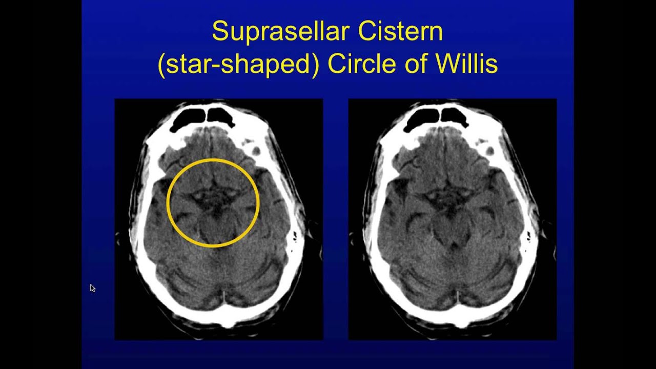
Basal subarachnoid cisterns normal configuration. It is sometimes defined as including the quadrigerminal cistern. Non contrast coronal ct head.
Radiology basics - Head anatomy. The procedure creates medical images of certain parts of the brain including the cerebrum brainstem and cerebellum. Anatomy of the head on a cranial CT Scan.
 058t 2 Gif 512 512 Radiology Imaging Pet Ct Brain Anatomy
 Subarachnoid Hemorrhage Ct Radiology Imaging Neurology Radiology
 Coronal Brain Ct Lateral Ventricle Cerebral Falx Brain Images Anatomy Interactive Anatomy
 Anatomy Of Head Ct Scan Normal The Brain On Ct And Mri With A Few Brain Images Ct Scan Brain Anatomy
 Ct Brain Anatomy Basal Ganglia Google Search Brain Anatomy Mri Brain Radiology Imaging
 A Anterior Horn Of The Lateral Ventricle B Caudate Nucleus C Anterior Limb Of The Internal Capsule D Putamen And Globu Ct Scan Caudate Nucleus Brain
 Cross Sectional Anatomy Mri Brain 15 Jpg 800 578 Brain Anatomy Mri Brain Radiology
 Brain Mri Sectional View Introduction Www Anatomynote Com Mri Brain Brain Anatomy Mri
Cross Sectional Anatomy Of The Brain Mri Interventional Radiology Brain
 The Radiology Assistant Brain Anatomy Brain Anatomy Nerve Anatomy Cranial Nerves Anatomy
 Image Result For Ct Brain Anatomy Basal Ganglia Brain Anatomy Basal Ganglia Anatomy
 Image Result For Ct Brain Anatomy Basal Ganglia Brain Anatomy Basal Ganglia Anatomy
 Brain Ct Anatomy Cerebral Lobes Ventricles Brain Anatomy Anatomy Human Anatomy Chart
 Axial View Of A Head Computed Tomography Ct Scan Of Pineal Gland Calcification In The Very Center Of The Brain Fluori Brain Anatomy Ct Scan Radiology Imaging
 Normal Brain Anatomy As Demonstrated By Computerized Tomography Ct Scanning Brain Anatomy Computerized Tomography Neurology
 Sagittal Ct Head Corpus Callosum Subarachnoid Cisterns Brain Images Anatomy Interactive Anatomy
 Bones Of Cranium Frontal Bone Parietal Bone Calvaria Diploe Brain Images Interactive Anatomy Human Anatomy Chart
 Mri Sectional Anatomy Of Brain Brain Anatomy Mri Brain Mri
 The Radiology Assistant Brain Anatomy Brain Anatomy Diagnostic Imaging Anatomy
Post a Comment for "Ct Anatomy Of Brain"