Sectional Anatomy Of The Eye (superior View)
You may also find RPE cones rods ganglion cells horizontal cells amacrine cells as well. Superior to the jugular foramen is the internal acoustic meatus which is located in the anterior medial section of the petrous portion of the temporal bone and allows both the facial and vestibulocochlear nerve to leave the skull.
 Mri Neck Anatomy Free Mri Axial Neck Cross Sectional Anatomy Anatomy Images Radiology Radiology Imaging
Extrinsic muscles of eyeball Fascial sheath of eyeball Tenon.
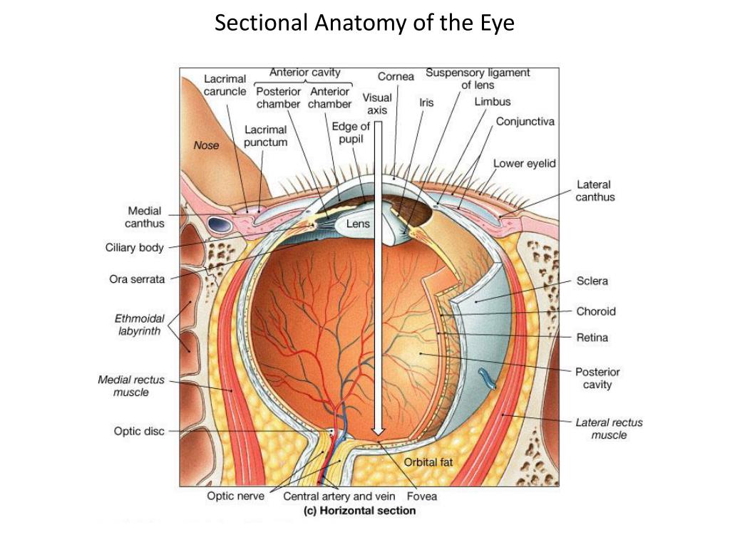
Sectional anatomy of the eye (superior view). Tough white colored fibrous portion of the outer layer of the eye. Light passes through the front of the eye cornea to the lens. Provides shape to the eyeball and attachment of muscles of the eye.
Composed of the MAXILLA Anterior portion and PALATINE Posterior portion bones. There are four rectus muscles medial lateral superior inferior and two oblique muscles superior and inferior when disease weakens or shortens these muscles eyes may go out of alignment and move poorly. The intermediate layer divided into two parts.
Filled with clear gel called vitreous humor. We hope this picture Anatomy of human eyes sectional view diagram can help you study. The central artery supplies the retina while the central vein drains the retina.
Anatomy of human eyes sectional view diagram. In the diagram above - anatomy of the eye the artery is shown in red while the vein is shown in blue. The external layer formed by the sclera and cornea.
We are pleased to provide you with the picture named Anatomy of human eyes sectional view diagram. Sagittal section of the adult human eye. This is an online quiz called Superior view of transverse section of right eye.
Anatomy of the eye. Anterior and posterior ciliary arteries. One portion is in a shallow depression in the part of the eye socket formed by the frontal bone.
The eye is kept moist by secretions of the lacrimal glands tear glands. One of six muscles that move eye. At Whitnall ligament it splits into the levator aponeurosis blue and the superior tarsal Müller muscle green which inserts at the superior border of the tarsus.
The eye works much the same as a camera. Both these nerves arise directly next to one another from the pontomedullary angle. Each gland has two portions.
The cells in the retina absorb and convert the light to electrochemical impulses which are transferred along the optic nerve and then to the brain. Illustrations Common tendinous ring. There is a printable worksheet available for download here so you can take the quiz with pen and paper.
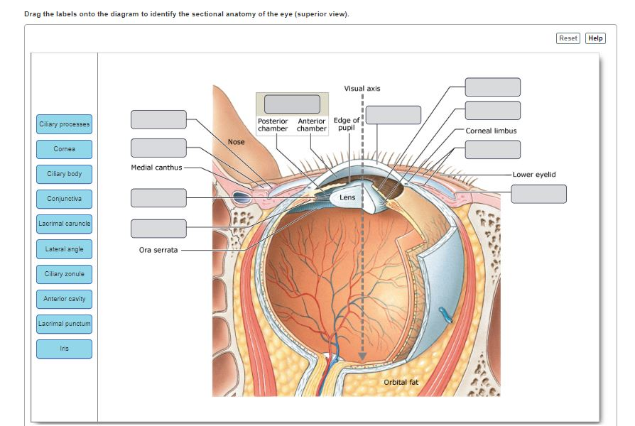
The orbit is open anteriorly where it is bound by the orbital septum which forms part of the eyelidsPosteriorly the orbit angles inward such that their apices communicate with the intracranial compartment via the optic canal and. The Sectional Anatomy of the Eye Superior View Part 1 Part A Drag the labels onto the diagram to identify the sectional anatomy of the eye superior view. Anterior iris and ciliary body and posterior choroid The internal layer or the sensory part of the eye the retina.
The Sectional Anatomy of the Eye Superior View Part 2 Visual axis Posterior Anterior Edge of chamber chamber pupil Cornea limbus Conjunt Nose Medial canthus -Lower eyelid purchim Lans Come Ora serrata Cary process Carybody Laura carundo Cilaryo Orbital fat 16. The superior oblique muscle or obliquus oculi superior is a fusiform muscle originating in the upper medial side of the orbit ie. Common anular tendon Zinn.
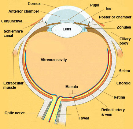
In this image you will find sclera cornea pupil lens iris ciliary body optic nerve fovea retina choroid in it. A cross-sectional view of the eye shows. Reset Help Visual axis Fovea centralis Nose Lateral cus Optic disc Lens Choroid i nono Posterior cart Medialrcius Reina Central artery and voin Sciara.
The central artery and vein runs through the center of the optic nerve. From beside the nose which abducts depresses and internally rotates the eye. The levator palpebrae superioris red has firm attachments to anterior aspect of the tarsus approximately 3 mm superior to the eyelid margin.
Identify the structure of the eye at the end of the red arrow. Drag the labels onto the diagram to identify the sectional anatomy of the eye superior view. The Sectional View Of The Eye Anatomy The Sectional View Of The Eye Anatomy In this image you will find the ciliary body iris cornea tear film pupil crystalline lens vitreous gel optic nerve macula fovea the retina in it.
In the adult the orbit has a volume of approximately 30 mL of which the globe occupies 65 mL. It has a roof floor medial and lateral wall. Composed of the vomer and perpendicular plate of ethmoid.
It is the only extraocular muscle innervated by the trochlear nerve the fourth cranial nerve. These almond-shaped glands under the upper lids extend inward from the outer corner of each eye. Its 25 anterior and superior to the EAM.
Houses the Sella Turcia which holds the pituitary gland. The cornea and the lens help to focus the light rays onto the back of the eye retina.
 The Sectional Anatomy Of The Eye Page 1 Line 17qq Com
 Pin On Be Safe At Night Sleep With A Nurse
 Cross Sectional Diagram In Which The Principal Structures Of The Midbrain At The Level Of The Superior Colliculus Are Brain Anatomy Anatomy Human Body Anatomy
 Solved Drag The Labels Onto The Diagram To Identify The S Chegg Com
 Cross Sectional Anatomy Of The Upper And Lower Eyelids Download Scientific Diagram
 Mri Anatomy Brain Axial Image 9 Brain Anatomy Mri Brain Anatomy
 18 21 E Sectional Anatomy Of The Eye And Accessory Structures Diagram Quizlet
Ppt Photoreception Vision Powerpoint Presentation Free Download Id 3644020
 F880d64511322a8d3c2764844ba60a35 Jpg 736 531 Brain Anatomy Mri Brain Radiology
 A P2 Lab 6 Hw Flashcards Quizlet
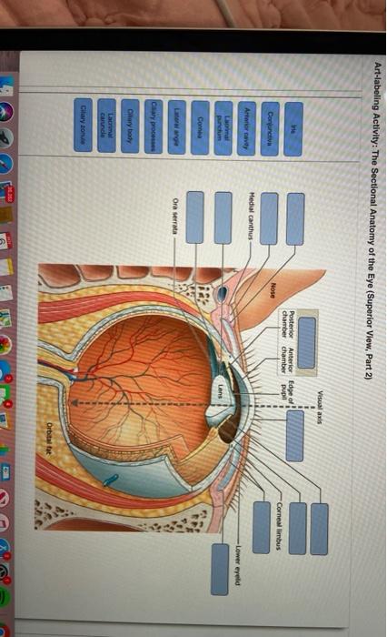
 Figure 17 5b The Sectional Anatomy Of The Eye Ppt Download
 Body Section Anatomy Anatomy Drawing Diagram
 Sectional Anatomy Of The Eye Page 1 Line 17qq Com
 Eye Cross Section Anatomy With English Name Stock Illustration 53060293 Pixta
 Skull Sectional View Anatomy Medical Anatomy Skull Anatomy
 Ct Cross Sectional Anatomy Of The Thoracic Cavity Arteries And Veins Vena Cava Heart Anatomy
 Cross Sectional Muscle Arrangement Around The Humerus Just Inferior To The Deltoid Muscle Attachment Anatomy Human Anatomy Cross Section
Post a Comment for "Sectional Anatomy Of The Eye (superior View)"