Structures That Protect The Eye
Answered Dec 14 2016 by GMCMaster. This might seem like an unnecessary precaution but it is okay to look geeky and protect your eyes from an injury especially if you are doing something that you are not trained to do.
 Fascinating Facts 20 Eye Opening Eye Facts Eye Anatomy Body Anatomy Human Body Anatomy
It is a white visible portion.

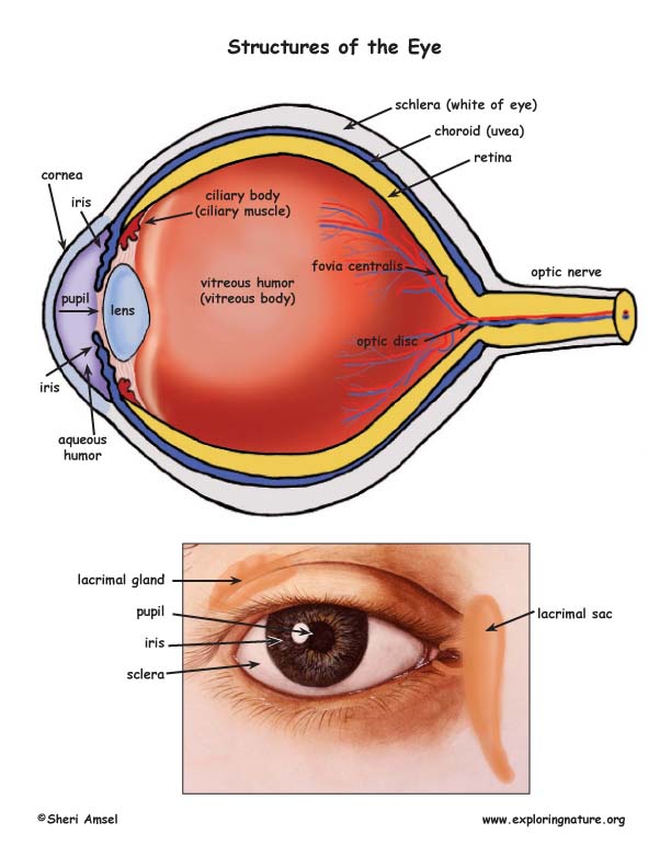
Structures that protect the eye. These accessory organs include the eyelids and lacrimal apparatus which protect the eye and a set of extrinsic muscles which move the eye. Spongy tissue located near the cornea through which aqueous humor flows out of the eye. Structures That Protect the Eye The bony structures of the orbit the bony cavity that contains the eyeball and its muscles nerves and blood vessels as well as the structures that produce and drain tears protrude beyond the surface of the eye.
Additional protection and support for the eye. The is a transparent tissue that bends and focuses light before it enters the lens. The eyeball is rounded so the cornea acts as a lens.
It keeps our eyes moist and clear and provides lubrication by secreting mucus and tears. The eye is a delicate organ which is protected by several structures such as eyebrows eyelids eyelashes and extraocular muscles. Accessory Structures of the Eye Accessory structures protect lubricate and move the eye.
Remove Contact Lenses Before Sleep. Eyebrows are two arched ridges of the supraorbital margins of the frontal bone. Is the transparent curved front of the eye which helps to converge the light rays which enter the eye Sclera Is an opaque fibrous protective outer structure.
The human eyelid features a row of eyelashes along the eyelid margin which serve to heighten the protection of the eye from dust and foreign debris as well as from. To understand how the eye sees it helps to know the eye structures and functions. This can be either voluntarily or involuntarily.
It lines the sclera and is made up of stratified squamous epithelium. An eyelid is a thin fold of skin that covers and protects an eyeThe levator palpebrae superioris muscle retracts the eyelid exposing the cornea to the outside giving vision. Asked Dec 14 2016 in Health Professions by Lareinalinda.
The eye lacrimal gland and associated extrinsic muscles are housed in the orbital cavity or orbit of the skull. It is soft connective tissue and the spherical shape of the eye is maintained by the pressure of the liquid inside. The sclera is the outermost layer of tissue also called the white of the eye.
The SKLEHR-uh the tough white part of the eye is composed of tough fibrous tissue that protects the inner layers of the eye and supports and shapes the eyeball. The orbit eyelashes eyelids conjunctiva and lacrimal glands help protect the eyes. Eye Structure and Function.
The eye consists of three layers of tissue which make up the wall of the eye. To protect your eyes from irritants and other possible eye injuries it is important to wear safety goggles. It bends or refracts light.
The parts of the eye that are visible externally include the following-Sclera. This layer is a very stable fibrous membrane that continues to retain the shape of the eye and provides protection. The orbit eyelashes eyelids conjunctiva and lacrimal glands help protect the eyes.
It provides attachment surfaces for eye muscles Choroid. The most important of these are the eyelids two folds of skin and tissue upper and lower that can be closed by means of muscles to form a protective covering over the eyeball against excessive light and mechanical injury. List the four structures that protect the eye eye lids eye lashes conjunctiva lacramal glands eye sockets and the bones of the skull Name the 5 parts of the eye that light rays pass through to focus on the retina.
The tough outer coat that protects the entire eyeball. The structures that protect the eyes are known as. It is made up of dense connective tissue and protects the inner parts.
Near the front of the eye in the area protected by the eyelids the sclera is covered by a thin transparent membrane conjunctiva which runs to the edge of the cornea. At the front of the eye is the cornea. Photoreceptor nerve cells in the eyes that are sensitive to low light levels and are present in the retina but outside the macula.
Connected to the sclera are the extra-ocular or extrinsic muscles of the eye. Several structures not parts of the eyeball contribute to the protection of the eye. Also absorbs stray light retina Receives the light and changes it into electrical impulses that travel to the brain.
Light enters through the cornea the transparent outer covering of the eye. They include the eyebrows eyelids conjunctiva lacrimal apparatus and extrinsic eye muscles figures 97 and 98. Structures That Protect the Eye The bony structures of the orbit the bony cavity that contains the eyeball and its muscles nerves and blood vessels as well as the structures that produce and drain tears protrude beyond the surface of the eye.
The conjunctiva also covers the moist back surface of the eyelids and eyeballs.
 Eye And Ear Models Eye Anatomy Anatomy Models Medical Anatomy
 Structures Of Eye Medical Medical School Studying Medical Dictionary
 Eye Anatomy Medical Knowledge Eye Anatomy Human Anatomy And Physiology
 Learn About The Human Eye Science For Kids Medical Anatomy Human Anatomy And Physiology Eye Anatomy
 Veterinary Online Ophthalmology Animals Veterinary Online Eye Anatomy Eye Anatomy Diagram Medical Anatomy
 Human Eye Anatomy Parts Of The Eye Explained Eye Anatomy Anatomy Human Anatomy And Physiology
Exploring The World Of Vision A Little Bit Of Basic Ocular Eye Health Homeopathy Treatment Medical Knowledge
 Human Eye Eye Anatomy Biology Diagrams Eye Study
 Landa Landa Eye Care Specialists Llc Eye Care Human Anatomy And Physiology Eye Anatomy
 Image Result For Kids Anatomy Chart Human Eye Diagram Eyeball Diagram Diagram Of The Eye
 Eye Anatomy And Eye Diagram Iris Pharma Eye Anatomy Eye Anatomy Diagram Anatomy
 Eye Anatomy 1 Illustration Photo In 2021 Eye Anatomy Eye Anatomy Diagram Human Anatomy And Physiology
 The Eye Diagram And Functions Functions Of The Human Eye Anatomy Body System Human Eye Diagram Eye Anatomy Diagram Of The Eye
 Vision And The Structure Of The Eye
 Major Ocular Structures Laramy K Independent Optical Lab Freeform Lenses And Ar Coatings
 Health Problem Solutions Accessory Structures Of The Eye Eye Anatomy Human Anatomy And Physiology Anatomy
 Conduct Eye Skin Care Medical Mnemonics Eye Facts
 All About The Structure Of The Human Eye Optimax
 Pin On Holding
Post a Comment for "Structures That Protect The Eye"