Anatomy Of Foot Joints
A joint is an articulation between two bones in the body and are broadly classified by the tissue which connects the bones. These joints form the ball of the foot.
 The Bones In The Foot Inferior View Picture Illustrated From Thieme Atlas Of Anatomy General Anatomy And Musc Anatomy Bones Human Bones Anatomy Body Anatomy
Fibrous joints have fibrous tissue joining the bone and these joints are typically very strong.

Anatomy of foot joints. While it has several more joints than the hind-foot it still possesses little mobility. These joints are the subtalar talocalcaneal talocalcaneonavicular calcaneocuboid cuneonavicular cuboideonavicular and intercuneiform joints. Also for students and health professionals it is critical to understand the foot anatomy which basically improves your knowledge of bone position ligament attachment and the way tendons run on the foot bones.
Lateral malleolus of the fibula. The cuneiform bones the navicularis and the cuboid all of which function to give your foot a solid yet somewhat flexible foundation. - Transection across the transverse tarsal joint is a standard method for surgical amputation of the foot.
The talus which is the bone in your ankle. Describe the components function of the foot ankle joints. The anatomy of the foot.
Tarsals five irregularly shaped bones of the midfoot that form the foots arch. The joints of the foot contain intertarsal tarsometatarsal intermetatarsal metatarsophalangeal and interphalangeal joints. The ankle joint also known as the talocrural joint is a hinge joint that involves the tibia and fibula of the leg and the talus of the foot.
The metatarsal phalangeal joints are the joints between the metatarsals and the proximal phalanx of each toe. This mortise is formed by the. The foot is divided into 3 categories.
Intertarsal tarsometatarsal metatarsophalangeal and interphalangeal. The three main types of joints are. The metatarsals articulate with the mid-foot at their base a joint called the tarsal-metatarsal TMT joint or Lisfranc joint.
The anatomic structures below the ankle joint comprise the foot which includes. The foot is divided into three sections - the forefoot the midfoot and the hindfoot. This consists of five long metatarsal bones and five shorter bones that form the toes phalanges.
Calcaneus the largest bone of the foot which lies beneath the talus to form the heel bone. The tarsal bones are the cuboid navicular and medial intermediate and lateral cuneiforms. The foot is made up of 26 bones 33 joints and over 100 ligaments tendon and muscle.
The calcaneus which is the bone in your heel. An example of which would be the sutures joining the various bones of the skull forming immovable joints. Articular surfaces fibrous capsule synovial membrane Ligaments medialdeltoid lateraltri-fascicular Movements plantardorsi flexion subtalar joints.
Foot and ankle anatomy is quite complex. Hindfoot the most posterior aspect of the foot is composed of the talus and calcaneus two of the seven tarsal bones. Mid-foot The midfoot begins with the calcaneal-cuboid joint and essentially ends where the metatarsals begin.
Medial malleolus of the tibia. Each big toe has two joints the metatarsophalangeal joint and the interphalangeal joint. The first three metatarsals medially are more rigidly held in place than the lateral two.
These all work together to bear weight allow movement and provide a stable base for us to stand and move on. It begins at the ankle joint and stops at the calcaneal-cuboid joint. Those joints that are of more functional worth are discussed.
These joints form the ball of the foot. Talus the bone on top of the foot that forms a joint with the two bones of the lower leg the tibia and fibula. This thickly laminated anatomical chart is printed on premium glossy 200 g UV resistant paper and comes with 2 sided lamination 125 micron 50 Mil and metal eyelets to make the chart easy to display.
The intertarsal joints are between the tarsal bones. The TMT joint is made stable not only by strong ligaments connecting these bones but also because the second metatarsal is recessed into the middle cuneiform in comparison to the others Figure 7. The metatarsophalangeal joint at the base of the toe the proximal interphalangeal joint in the middle of the toe and the distal phalangeal jointthe joint closest to the tip of the toe.
The foot contains a lot of moving parts - 26 bones 33 joints and over 100 ligaments. The fore foot metatarsals and phalanges mid foot cuboid navicular and 3 cuneiforms and hind foot talus and calcaneous. The talus and calcaneus articulation is referred to as the subtalar joint which has three facets on each of the talus and calcaneus.
Synovial cartilaginous and fibrous. The metatarsals which run through the flat part of your foot. Besides the ankle joint which connects the foot with the leg the bones of the foot articulate among themselves through many synovial joints.
Joints of the foot. There are four groups of foot joints. The feet are divided into three.
The poster also details some common pathologies of the foot and foot joints. The other four toes on each foot have three joints each. Fore-foot the fore-foot is composed of the metatarsals and phalanges.
The body of the talus sits within a deep recess referred to as the mortise. Hind foot fore foot mid foot fore foot mid foot hind foot Tarsometatarsal joint Transverse tarsal joint Subtalar joint Note. What you will learn in the article.
Each foot contains five metatarsals numbered 1-5 medial great toe to lateral. Distal tibiofibular joint talo-calcaneo-navicular mid-tarsal joint. The foot consists of thirty three bones twenty six joints and over a hundred muscles ligaments and tendons.
The feet are flexible structures of bones joints muscles and soft tissues that let us stand upright and perform activities like walking running and jumping. There are numerous joints in the foot created between tarsal metatarsal and phalangeal bones. - The complex anatomy of the Chopart joint optimally adapting to uneven surfaces upon first heel contact forcefully pushing the foot off the ground at the end of the walk cycle.
The joints will be discussed later in the tutorial. This colorful anatomical poster illustrates the anatomy of the foot and joints of the foot.
 Foot Anatomy Bones Bottom View Anatomy Bones Foot Anatomy Bones Skeleton Anatomy
 Foot Bone Anatomy Overview Tarsal Bones Gross Anatomy Metatarsal Bones Gross Anatomy Body Diagram Foot Bone Anatomy Human Bones
 Bones Of The Right Foot Human Body Anatomy Anatomy Bones Body Anatomy
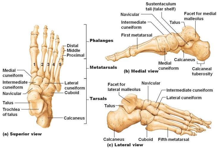
 Foot Bones Top View Anatomy Bones Foot Anatomy Human Anatomy
 Anatomy Of Foot Bones Anatomy Amp Physiology 141 Gt Ganther Gt Flashcards Gt Chapt 6 Bones A Amp P Foot Anatomy Anatomy Bones Ankle Anatomy
 The Body Fig 12 32 Bones Of The Right Foot Click For High Resolution Image Anatomy Bones Human Anatomy Art Foot Anatomy Bones
 Foot Anatomy 01 Anatomy Bones Joints Anatomy Foot Anatomy
 Pin On Anatomy
 Foot Bone Anatomy On Healthfavo Com Health Medicine And Anatomy Reference Pictures Anatomy Bones Foot Bone Anatomy Anatomy Reference
 Drawing To Show The Bones Of The Right Foot Dorsal Or Top View Anatomy Bones Ankle Anatomy Human Anatomy And Physiology
Appendicular Skeleton Anatomy Bones Medical Technology Human Anatomy And Physiology
 List Of All Metatarsus Bones Check Out More Information Of Foot Anatomy Bones Http Www Learnbones Com Foot Bo Anatomy Bones Medical Anatomy Anatomy Organs
 Pin On Neat Feet
 Foot Anatomy Eorthopod Com Foot Anatomy Bone And Joint Medical Knowledge
 There Are 26 Bones In The Human Foot Which Are Grouped Into 7 Tarsals 5 Metatarsals And 14 Phalanges For Medical Anatomy Human Anatomy Anatomy And Physiology
 Lateral Aspect Of The Ankle Ligaments Netter Ankle Anatomy Muscle Anatomy Human Anatomy And Physiology
 Anatomy Of The Human Foot Koibana Info Foot Anatomy Anatomy Bones Human Anatomy Picture
 Anatomy Of The Foot Part 1 Bones And Joints Ankle Anatomy Anatomy Bones Anatomy Images
 Dorsum Of Foot Anatomy Bones Skeletal System Joints Of Foot Muscles Attachment Tarsal Bones Talus Calc Anatomy Bones Foot Anatomy Human Body Anatomy
Post a Comment for "Anatomy Of Foot Joints"